Equine Diagnostic Imaging
We use electromagnetic radiation and other technologies for diagnostic imaging. This allows us to produce highly detailed images of your pet's internal structures.
At Pacific Crest Equine, we have advanced tools to help diagnose your pet's medical issues.
We offer a variety of services, from digital radiology to ultrasound and videoendoscopy.
With our diagnostic imaging capabilities, we can efficiently produce diagnostic information about your pet's condition and provide immediate treatment options.

Field Services for Horses Throughout the San Joaquin Valley
If your horse needs veterinary care at your farm, our veterinarians can come to you with fully equipped trucks, stocked with the technology and tools needed to diagnose your horse's condition or provide routine veterinary care.

Our Imaging Capabilities
With our in-house veterinary diagnostics lab, we are pleased to offer advanced diagnostic testing to allow our vets to provide a diagnosis of your horses medical issues.
- Radiography (Digital X-rays)
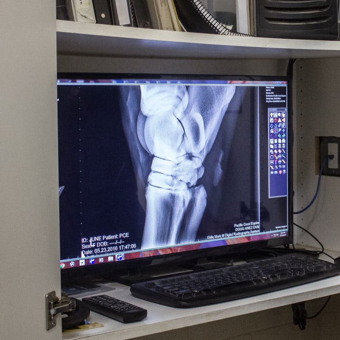
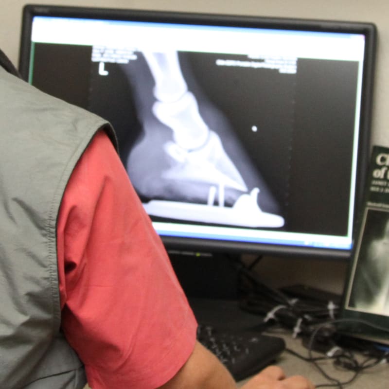
Radiographs are often the first diagnostics imaging modality used to evaluate lameness. Digital Radiography allows for potentially greater detail than conventional radiography by having each image be processed by the computer.
Digital radiography services for equine patients both in our hospital and on the farm allow us to diagnose issues such as fractures, laminitis, wounds, foreign objects, and penetration of joints. They can also be used for pre-purchase exams and lameness evaluations.
- Ultrasound
Ultrasound uses sound waves to offer a non-invasive, painless, and radiation-free method of imaging your horse's internal organs, tendons, and ligaments. Our hospital is equipped with a state-of-the-art ultrasound with superior image quality and detail, which allows us to evaluate tendons and ligaments with the utmost precision and accuracy.
Small ligament tears and suspensory ligament injuries can be diagnosed quickly and we can scan abdomens, perform cardiac evaluations, and diagnose pelvic and back muscle pathology with this system. Images are recorded and stored digitally and therefore can be cataloged and viewed sequentially for each patient.
One of the best features of this machine is its capability for quantitative analysis for tendon measuring. The software can analyze the images produced from scans to produce and injury score. This enables us to give the horse a more accurate diagnosis, the prognosis for return to work, and an individualized treatment plan.
Ultrasound can also be used to guide technical joint injections such as cervical spine (neck), lumbar spine (low back), and the sacroiliac joint (SI). Ultrasound is also useful in diagnosing masses or lumps. This can be used not only externally but internally as well.
- Videoendoscopy
A videoendoscope is used for viewing and evaluating the equine upper airway, the esophagus, the stomach, and many components of the reproductive tract and bladder. This provides a higher-quality image than older technology that used fiber optic endoscopes. In addition, both still photographs and video images can be captured and recorded for future review.
We have both one- and three-meter video endoscopes available to suit our equine patient's individual needs.
- Thermography
Our FLIR thermography system is available to scan horses experiencing a variety of problems such as lameness or back soreness, poor saddle fit, and shoeing and hoof balance problems.
This technology can be used to scan horses for areas of increased heat and inflammation or decreased temperature from pathology and disuse. Thermography can be used to locate soft tissue injuries often before they can be seen on physical examinations or ones that cannot be seen on radiographs.

